Canon Products
CX-1
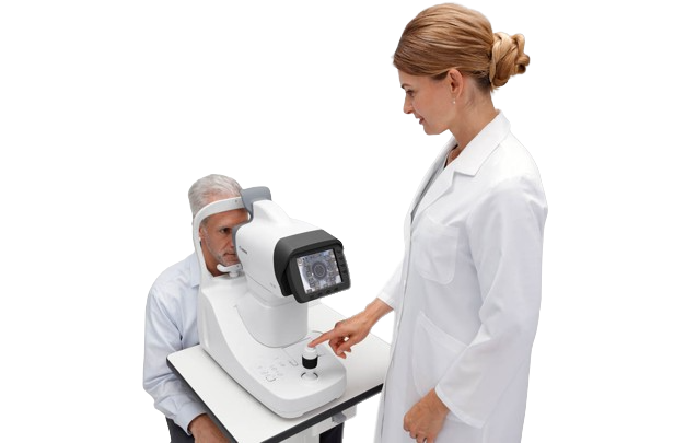
Hybrid Digital Mydriatic/Non-Mydriatic Retinal Camera

Hybrid Digital Mydriatic/Non-Mydriatic Retinal Camera
CX-1
The CX-1 Hybrid Digital Mydriatic/Non-Mydriatic (MYD/NM) Retinal Camera is Canon's first hybrid camera and a non-mydriatic camera to use Fundus Autofluorescence (FAF) photography.
Five Photography Modes
The CX-1 offers five photography modes including Color, Red-Free, Cobalt, Fluorescein Angiography (FA), and Fundus Autofluorescence (FAF).


Non-Mydriatic Fundus Autofluorescence (FAF) Photography
With the push of the Myd/Non-Myd button to select the non-mydriatic mode and the FAF mode, the operator is ready to begin taking images. The FAF mode helps monitor macular waste such as lipofuscin, which assists the eye care professional with detecting age-macular degeneration (AMD), glaucoma, and diabetes.


One Touch Selection of MYD/NM
A single push of the Myd/Non-Myd button switches between the mydriatic and non-mydriatic imaging modes.

Control Panel
Whether the CX-1 is in mydriatic or non-mydriatic mode, the control panel has simple operation, workflow efficiency, and ergonomic controls.

Superior Image Quality
Clear, 32.5 megapixel high resolution images are available in either mydriatic or non-mydriatic mode. The 2x mode magnifies the image by automatically cropping out the peripheral edges so the region of interest is larger in the frame.

Stereo Photography System
The LCD monitor displays guides which automatically determine the base length for acquiring successful stereo images. The two images can be stored as a pair making it easier to manage the files.

EOS Camera Technology
Made exclusively for the CX-1, the detachable camera features Canon's EOS camera technology with its renowned image processing capabilities providing optimal retinal imaging. The camera handles five different photography modes including non-mydriatic FAF photography, Color, Red-free, Fluorescein Angiography, and Cobalt.

CR-2 AF
Digital Non-Mydriatic Retinal Camera

Digital Non-Mydriatic Retinal Camera
CR-2 AF
The CR-2 AF Digital Non-Mydriatic Retinal Camera features the latest in Canon retinal imaging technology and enhancements in a compact and light weight design. The CR-2 AF also provides autofunctionality with contrast enhancement. It can be installed and takes up minimal space. For added convenience, the CR-2 AF camera can remain mobile for easy transportation when needed using an optional hard shell transport case, sold separately

Auto-Focus with Manual Alignment Override
Automatically focuses the eye by partially depressing the joystick, or easily switches to manual focus with a twist of the focus ring.
Auto-Capture
Automatically captures the image once the eye is properly focused.
Auto-Exposure
Automatically measures the volume of infrared light at the retina and adjusts the flash intensity.
Auto-Fundus
Automatically switches from the external eye to retinal observation mode when the eye is properly aligned.
Image Error Detection
Advanced software automatically confirms both correct alignment and focus.
Contrast Enhancement
Provides increased image clarity by emphasizing the difference in 'redness' and 'brightness' of blood vessel structures relative to their surroundings.

High Resolution Images
The CR-2 AF provides ultra-high resolution, 32.5 megapixel, wide-angle views with excellent color, detail and contrast, making it easy for the diagnosis and detection of ocular conditions and diseases.

Digital Filter Processing
With digital red-free, digital cobalt mode, the CR-2 AF extracts each element from the color image. Additional filter view are available with imageSPECTRUM Image Management System software (sold separately).


Quick Preview
Preview images directly on the dedicated EOS camera immediately after capture.

Low Flash Intensity
Low flash improves patient comfort and reduces miosis for a faster exam. The CR-2 AF supports a wide range of low ISO speeds, including ISO 200, 400, 800, 1600, 3200, and 6400.

CR-2 PLUS AF
Digital Non-Mydriatic Retinal Camera

Digital Non-Mydriatic Retinal Camera
CR-2 PLUS AF
The Canon CR-2 PLUS AF Digital Non-Mydriatic Retinal Camera provides Color and Fundus Autofluorescence (FAF) imaging within a small compact design. Features include Auto-Fundus, Auto-Focus, Auto-Capture and Image Error Detection. Geographic Atrophy, Macular Degeneration, Glaucoma, Diabetic Retinopathy and other conditions that can affect vision may also be identified and monitored using FAF mode. Using invisible infrared alignment light, the digital non-mydriatic camera may image patients with pupils as small as 3.3 mm (small pupil mode) without dilation drops. This is especially useful when performing retinal screenings or expediting routine retinal imaging exams during office visits.

Auto-Focus with Manual Alignment Override
Automatically focuses the eye by partially depressing the joystick, or easily switches to manual focus with a twist of the focus ring.
Auto-Capture
Automatically captures the image once the eye is properly focused.
Auto-Exposure
Automatically measures the volume of infrared light at the retina and adjusts the flash intensity.
Auto-Fundus
Automatically switches from the external eye to retinal observation mode when the eye is properly aligned.
Image Error Detection
Advanced software automatically confirms both correct alignment and focus.
Contrast Enhancement
Provides increased image clarity by emphasizing the difference in 'redness' and 'brightness' of blood vessel structures relative to their surroundings.
High Resolution Images
The CR-2 PLUS AF provides ultra-high resolution, 32.5 megapixel, wide-angle views with excellent color, detail and contrast, making it easy for the diagnosis and detection of ocular conditions and diseases.
Fundus Autofluorescence (FAF)
The CR-2 PLUS AF Digital Retinal Camera model enables you to assess and monitor the condition of the Retinal Pigment Epithelium (RPE) using the Fundus Autofluorescence (FAF) imaging mode.

Digital Filter Processing
With digital red-free, digital cobalt mode, the CR-2 PLUS AF extracts each element from the color image. Additional filter view are available with imageSPECTRUM Image Management System software (sold separately).
Quick Preview
Preview images directly on the dedicated EOS camera immediately after capture.
Low Flash Intensity
Low flash improves patient comfort and reduces miosis for a faster exam. The CR-2 PLUS AF supports a wide range of low ISO speeds, including ISO 200, 400, 800, 1600, 3200, and 6400.
TX-20
Full Auto Tonometer

Full Auto Tonometer
TX-20
The Canon TX-20 Full Auto Tonometer easily measures Intraocular Pressure (IOP) allowing doctors to perform simple eye exams efficiently while enhancing patient comfort with a soft air puff. This test is important in evaluating ocular conditions that affect the pressure inside of an eye, such as Glaucoma. Whenever high IOP is detected, a warning message is displayed. The operator can enter any threshold value on the unit's settings screen to activate the warning system for that value. The TX-20 Tonometer is compact and lightweight at approximately 33 pounds, making it easy to transport and install.

Selectable Measurement Modes
The three-dimensional tracking system of the TX-20 Tonometer features a wide range of movement, so that the examinee's pupil can be detected easily. When in Full Auto mode, the TX-20 Tonometer is a one touch operation for IOP measurement for both eyes. Just press the start button and the unit automatically aligns and measures the intraocular pressure of both eyes. For added flexibility, the TX-20 Tonometer also includes Auto and Manual measurement modes.
Soft Air Puff & Position Safety Alert
For many patients, the only thing significant and worrisome about tonometry is the air puff. The TX-20 Tonometer employs a delicate air puff for patient comfort. When the Safety Alert is set, the TX-20 Tonometer automatically prevents the optical head from making physical contact with the patient.
Visual Reliability & Indicators
When a reading cannot be made, the TX-20 Tonometer display will show a snapshot of the examinee's eye along with the measurement error. The operator will know immediately whether it was an eyelid, eyelash or the eye being out of position that caused the error. The operator will then be informed on the next action to take such as adjusting the chin rest or limiter.
External Fixation
The external fixation is effective for patients with central vision defects and can be activated using the menus on the LCD screen.

Compact and Lightweight
Made for comfort for both the patient and the operator, the TX-20 Tonometer maximizes a small work area with its compact design. At approximately 33 pounds, it comfortably fits on an instrument table with a Canon Non-Mydriatic Retinal Camera (each sold separately.) Keeping the Canon machines next to one another can help to expedite the exam process.

Multifunctional Color LCD Monitor
The 5.7 inch color LCD monitor tilts 40° making the TX-20 Tonometer easy to use whether the operator is sitting or standing. The clear LCD display also includes multi-functional screen buttons allowing the operator to switch menus as needed.

Motorized Omni-Directional Joystick
Aligning patients is made easier using the motorized Omni-directional joystick with a fine focus dial. All positioning may be performed with one hand allowing the other hand to work with the patient, if needed. The top of the joystick button includes the start button, which can immediately be pressed once the patient is properly positioned.

Input & Output Modes
The TX-20 Tonometer provides data output using RS-232C and LAN connections, and data input using USB connections.

Printouts & Fast Built-In Printer
Internal memory provides storage of up to 10 measurements for each eye, listing in the order they were taken, or by reliability. A printout can be setup to automatically print. The front loaded built-in printer includes an auto cutter, making it simple to remove the print-out. New paper rolls (sold separately) can easily be inserted through the feeder.

Xephilio OCT-A1
Optical Coherence Tomography

Optical Coherence Tomography
Xephilio OCT-A1
The Xephilio OCT-A1 Optical Coherence Tomography is Canon's Optical Coherence Tomography for the in-vivo imaging and measurement of the retina, retinal nerve fiber layer, and optic disc.
.png)
High-Definition Imaging
The Xephilio OCT-A1 System, which offers 1.6 μm Axial digital resolution in combination with the ability to average multiple scans, can help provide excellent image quality with detailed resolution.

Accurate Scanning, Outstanding Ease of Use
The System’s integrated Scanning Laser Ophthalmoscope (SLO) contributes significantly to scan quality and ease of use. By providing real-time retinal tracking, it makes monitoring of the examination easy.

Fast and Precise Follow-up
The SLO also assists with followup examinations by automatically adjusting to the same scan position as used in the previous exam. To ease comparison, the software automatically selects identical scan parameters.

High Definition, Enhanced Depth, Wide Field of View
With the Xephilio OCT-A1 System, you can average up to 50 cross scans* to achieve an image resolution that allows impressive detail of both the layer and the vitreous pleated structures. For optimal imaging, the System offers special scan modes for vitreous and choroid imaging in addition to a wide scan width of up to 13 mm.

Reliable 10-Layer Recognition
The Xephilio OCT-A1 System can automatically detect and distinguish 10 layers of the retina— including Bruch’s membrane (BM)—thanks to its excellent image quality and resolution.

Xephilio OCT-S1
Wider and deeper, wide-field swept source imaging in one single capture
Canon introduces Xephilio OCT-S1, revolutionary swept source technology allowing wide-field retina imaging of up to 23mm in a single scan. Xephilio OCT-S1 enables superior penetration of dense objects and provides outstanding tomographic images. The system’s Deep Learning AI technology, Intelligent Denoise, offers a new quality of OCTA images in a single scan with greatly reduced noise, increased details and improved visibility within seconds.
- Wide-field imaging in one single capture
- AI Intelligent Denoise Processing Function
- Improved patient’s comfort


Features
Wide-field Imaging in One Single Capture
The Xephilio OCT-A1 Optical Coherence Tomography is Canon's Optical Coherence Tomography for the in-vivo imaging and measurement of the retina, retinal nerve fiber layer, and optic disc.

AI Intelligent Denoise Processing Function
With a single click of our Artificial Intelligent Noise Reduction Process (Intelligent Denoise), high quality images with low noise can be easily acquired. In addition, this process can be completed in a short time regardless of the size of the image, thereby greatly improved the overall efficiency.

Improved Patient’s Comfort
With Xephilio OCT-S1, wide-field images equaling approximately 80° viewing angle [horizontal 78°, vertical 68°, 8μm (typical)] and up to 5.3mm depth can be acquired in just one scan. Vascular abnormalities (non-perfusion, new blood vessels, etc. in the surrounding area with a single image such as diabetic retinopathy and arteriovenous obstruction. Compared to conventional panoramic photography, the strain on the patient is greatly reduced.

- Specifications, availability and terms of offers may change without notice.
- Products / Services may be manufactured by and/or supplied to us by third party manufacturers / suppliers for distribution / resale (non-Canon brand products).
- Please refer to individual country / region websites and respective sales offices for product availability.
RK-F3m
Designed for ease of use with
multiple useful modes, versatile and compact
One-touch smooth operation for accurate, automatic measurement of both eyes.
- Easy Operation with Large LCD Monitor
- Flexible Examination Layout
- Multiple Measurement Modes


Features
Easy Operation with Large LCD Monitor
10.4-inch touch panel colour LCD provides easy viewing of alignment and measurement results, making measurements a breeze.

Flexible Examination Layout
Tilt and rotatable LCD monitor, allowing flexibility in operation suitable for multiple examination layouts.

Multiple Measurement Modes
- Improved verification of refractive power and accommodation mode.
- Retroillumination mode with auto gain.
- Wide range of available VD values.

- Please refer to individual country / region websites and respective sales offices for product availability.
- Specifications, availability and terms of offers may change without notice.
- Products / Services may be manufactured by and/or supplied to us by third party manufacturers / suppliers for distribution / resale (non-Canon brand products).
APPASAMY ASSOCIATES (P) LTD,No.20, SBI Officer's colony,
1st Street, Arumbakkam, Chennai - 600 106, Tamil Nadu, India.
Call: +91 6382758855 / +91 9381216555
E-mail : info@appasamy.com
©2024 Appasamy Associates Private Limited
.png)
